AP Inter 1st Year Botany 4th Lesson Anatomy of Flowering Plants Questions and Answers
IV. Very Short Answer Questions
Question 1.
A transverse section of a plant shows conjoint, scattered vascular bundles surrounded by sclerenchymatous bundle sheath. What will you identify it as?
Answer:
Monocot stem
Question 2.
Which parts of the leaf surface reduce the rate of transpiration?
Answer:
The waxy cuticle on the leaf surface and the number and position of stomata on the leaf surface both reduce the rate of transpiration.
Question 3.
What are the cells that make the leaves curl in plants during water stress? Give an Example.
Answer:
Bulliform cells.
Ex: Monocot leaves (Grass).
Question 4.
How is the study of plant anatomy useful to us?
Answer:
- Anatomy helps in understanding internal structure or arrangement of various tissues of plants.
- Anatomy helps in understanding structural details of different organs of a plant, anatomical differences found in between different kinds of plants like monocots and dicots, and also helps in knowing adaptations found in plants to diverse environments
Question 5.
Name any two simple tissues found in the ground tissue system.
Answer:
Parenchyma and collenchyma are two simple tissues found in the ground tissue system of plants.
Question 6.
What are the shapes of guard cells in dicot leaf and monocot leaf respectively?
Answer:
- In Dicot leaf- guard cells are Kidney or bean-shaped.
- In Monocot leaves- guard cells are dumbbell-shaped.
Question 7.
What are casparian strips? In which part of the plant anatomy do you find them?
Answer:
Casparian strips are ring-like cell wall thickenings found in the endodermis of the roots of vascular plants.
Question 8.
Write three words to describe the vascular bundle of a dicot stem.
Answer:
Vascular bundles in dicot stem are: conjoint (xylem and phloem are arranged in the same radius), open (a strip of cambium is present in between xylem and phloem) and endarch (protoxylem is present towards the inside and metaxylem is present towards periphery).
V. Short Answer Questions
Question 1.
Write any four differences between monocot leaf and dicot leaf.
Answer:
| Monocot leaf | Dicot leaf |
| 1. Trichomes are absent. | 1. Trichomes are present. |
| 2. Stomata are equal on both the layers. | 2. Stomata are more in the lower epidermis. |
| 3 Bulliform cells are present in the upper epidermis. | 3. Bulliform cells are absent. |
| 4 Mesophyll consists of only spongy tissue. | 4. Mesophyll is differentiated into palisade and spongy tissues. |
| 5. Bundle sheath extensions are sclerenchymatous. | 5. Bundle sheath extensions are collenchymatous. |
| 6. Vascular bundles are similar in size. | 6. Vascular bundles are big in the mid vein. |
Question 2.
Cut a transverse section of the young stem of a plant from your garden and observe it under the microscope. How would you ascertain whether it is a monocot stem or a dicot stem? Give reasons.
Answer:
I) The given transverse section of the young stem can be ascertained as a dicot stem based on the following characters.
- The epidermis bear trichomes.
- Cortex is well distinguished into three subzones viz., collenchymatous hypodermis, general cortex and strachy endodermis.
- Pericycle is present as semilunar patches of sclerenchyma, between endodermis and phloem of vascular bundles.
- A large number of vascular bundles are arranged in a ring around the medulla.
- Vascular bundles are conjoint and open.
- Medulla (or) pith and Medullary rays are present.
II) The given transverse section of the young stem can be ascertained as monocot stem based on the following characters.
- Trichomes are absent in the epidermis.
- Cortex is highly reduced and represented by sclerenchyrhatous hypodermis.
- Endodermis and pericycle are absent.
- A large number of vascular bundles surrounded by a sclerenchymatous bundle sheath are scattered in the ground tissue.
- Vascular bundles are conjoint and closed and phloem parenchyma is absent. In the vascular bundles, water-containing lysigenous cavities are present.
- Peripheral vascular bundles are generally smaller than the centrally located ones.
- Medulla (or) pith and medullary rays are absent.
Question 3.
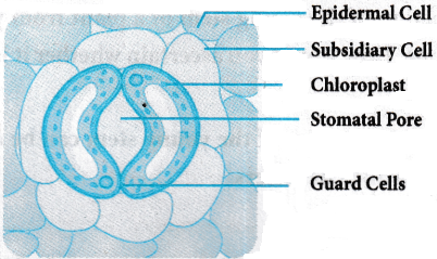
The stomatal pore is guarded by two kidney-shaped guard cells. Name the epidermal cells surrounding the guard cells. How does a guard cell differ from an epidermal cell? Use a diagram to illustrate your answer.
Answer:
-
- The stomatal pore is guarded by two kidney-shaped guard cells.
- These guard cells are surrounded by the epidermal cells known as subsidiary cells or accessory cells.
| Guard cells | Epidermal cells |
| 1. The guard cells are smaller in size, bean-shaped or dumbbell-shaped. | 1. They are barrel-shaped. |
| 2. Chloroplasts are present. | 2. Chloroplasts are absent. |
Question 4.
Name the three basic tissue systems in the flowering plants. Give the tissue names under each system.
Answer:
The three basic tissue systems of flowering plants are
a) Epidermal tissue system
b) Ground tissue system
c) Vascular tissue system
a) Epidermal tissue system:
It forms the outermost covering of the whole plant body and comprises Epidermis, Stomata, Epidermal appendages like the trichomes and root hairs.
b) Ground tissue system :
All tissue, except epidermis and vascular bundles constitute the ground tissues. It consists of hypodermis, general cortex, endodermis, pericycle, medulla and medullary rays.
c) Vascular tissue system:
- It consists of complex tissues like phloem and xylem, arranged in the form of bundles.
- Vascular Bundles are of Radial (Roots), Collateral (Stem) type.
VI. Long Answer Questions
Question 1.
Describe the internal structure of dorsiventral leaf with the help of labelled diagram.
Answer:
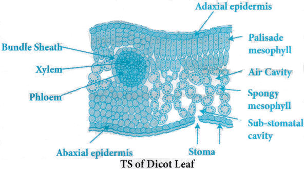
The transverse section of dorsiventral leaf (dicot leaf) shows three parts, namely
I) Epidermis,
II) Mesophyll,
III) Vascular bundle
I) Epidermis:
A single layered epidermis is present towards the lower (abaxial) and upper side (adaxial) of the leaf called lower epidermis and upper epidermis respectively.
Both adaxial and abaxial epidermis of leaves covered by a cuticle.
Stomata are more towards lower or abaxial epidermis.
II) Mesophyll:
Tissue present between upper and lower epidermis is called Mesophyll. It is of two kinds,
a) Palisade parenchyma and
b) Spongy parenchyma.
a) Palisade parenchyma:
- It is present below the upper epidermis that is towards the adaxial surface.
- It shows elongated cells which are arranged vertically and parallel to each other.
b) Spongy parenchyma:
- Part of the mesophyll towards the lower epidermis or abaxial surface is called spongy parenchyma.
- Spongy parenchyma is situated below the palisade cells and extends to the lower epidermis.
- It is made up of oval or round shaped cells with large intercellular spaces.
III) Vascular bundle :
- Vascular bundles are extended in the mesophyll in the form of vein and the midrib.
- The vascular bundles are conjoint, collateral and closed.
- The xylem is present towards the upper side (adaxial) and phloem is paresent towards lower epidermis (abaxial) in each vascular bundle.
- The vascular bundle is surrounded by a layer of thick-walled cells called bundle sheath. The cells are arranged compactly.
Question 2.
What is a stomatal apparatus? Explain the structure of stomata with a labelled diagram.
Answer:
- Stomata are tiny pores in the epidermis of leaves and other plant parts.
- The stomata consist of minute pores called stoma surrounded by a pair of guard cells.
- Stomata, open and close according to the turgidity of guard cells.
- The cell wall surrounding the pore is tough and flexible.
- Guard cells are bean-shaped and contain chloroplasts.
- They contain chlorophyll and capture light energy.
- The Guard cells are surrounded by subsidiary cells.
- They are the accessory cells to guard cells and are found in the epidermis of plants.
- The stomatal aperture, guard cells and the surrounding subsidiary cells are together called stomatal apparatus.
Question 3.
Describe the T.S. of a dicot stem with the help of neat labelled diagram ?
Answer:
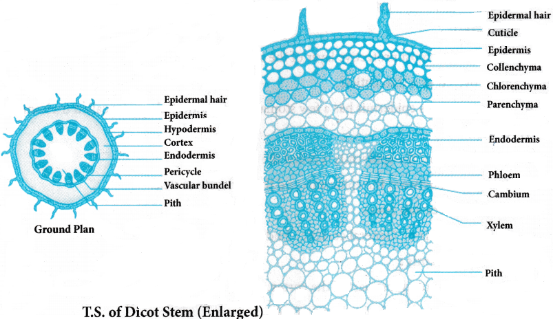
The transverse section of the young primary dicot stem is differentiated into
I) Epidermis
II) Cortex
III) Stele
I) Epidermis:
- It is the outermost protective layer of the stem.
- It is covered by a thin cuticle.
- In young stems epidermis has a few stomata and multicellular hairs called trichomes.
II) Cortex:
- The part below the epidermis is called the cortex.
- It is present between the epidermis and the pericycle of stele.
- It shows three sub zones.
- a) Hypodermis
- b) General cortex
- c) Endodermis
a) Hypodermis:
- It is the outermost part of the cortex.
- It is composed of a few layers of collenchymatous cells.
- It provides mechanical strength (tensile) to the young stem.
b) General Cortex:
- It is present between the hypodermis and endodermis.
- It is composed of thin walled, round parenchyma cells with intercellular spaces.
c) Endodermis:
- The innermost layer of the cortex is called endodermis.
- Due to storage of large quantities of starch in the cells of endodermis, it is also called as “Starch sheath”.
III) Stele:
- The central cylinder concerned with conduction of water and minerals is called stele.
- It is differentiated into
a) Pericycle
b) vascular bundles
c) Medulla (or) Pith.
a) Pericycle:
- It is the outermost layer of the stele.
- It is present in the form of semilunar patches of sclerenchyma in between endodermis and phloem of vascular bundles.
b) Vascular bundles:
- A large number of vascular bundles are arranged in the form of a ring.
- This arrangement is called Eustele.
- Vascular bundles are conjoint, collateral, endarch and open type.
c) Medulla or Pith:
It is composed of a large number of rounded parenchymatous cells having intercellular spaces.
d) Medullary rays:
In between the vascular bundles there are a few layers of radially placed living parenchyma cells, which constitute medullary rays.
Question 4.
Describe the T.S. of a monocot stem with the help of labelled diagram ?
Answer:
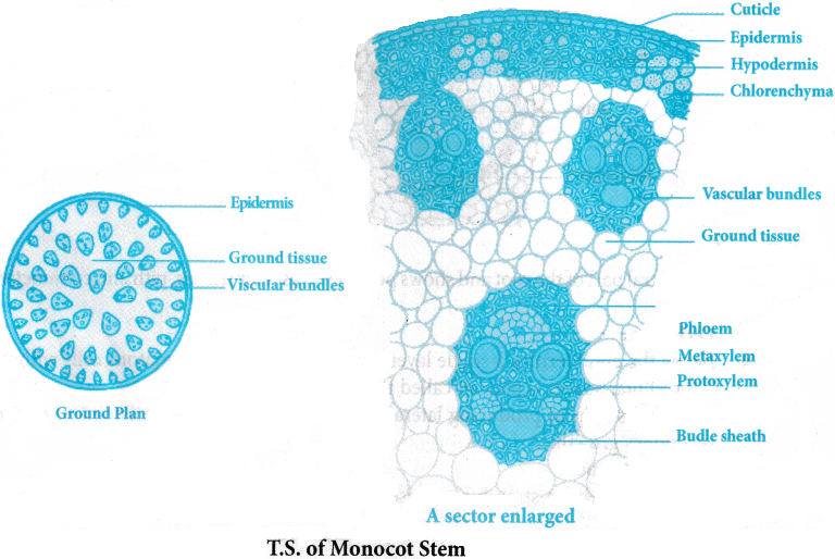
T.S of young monocotyledonous stem shows Epidermis, hypodermis, ground tissue and vascular bundles.
A) Epidermis:
- The outermost layer composed of compactly arranged cells.
- It is covered by a thin layer of cuticle.
- Trichomes are absent.
B) Hypodermis:
Below the epidermis, sclerenchymatous hypodermis is present, which gives mechanical support to the stem.
C) Ground tissue:
- Below the hypodermis, parenchymatous ground tissue is present with intercellular spaces.
- Endodermis and pericycle are absent.
D) Vascular bundles:
- A large number of vascular bundles are scattered in the ground tissue so called atactostele.
- Each vascular bundle is Collateral, conjoint, endarch and closed type.
- Each vascular bundle is surrounded by sclerenchymatous bundle sheath.
- The phloem parenchyma is absent and water containing cavities are present.
- Medulla and medullary rays are absent.
Question 5.
Describe the internal structure of a dicot root with the help of labelled diagram ?
Answer:
The internal structure of the dicot root shows Epidermis, cortex and stele.
A) Epidermis:
- The outermost layer is made up of compactly arranged elongated cells called Epiblema.
- Many of the cells produce unicellular root hairs which help in absorption of water from the soil.
B) Cortex:
- It is present in between the epidermis and the stele.
- Below the epidermis, several layered parenchymatous general cortex is present.
- The cells are thin walled living.
Endodermis:
- The innermost layer of the cortex is composed of a single layer of barrel shaped cells.
- The radial and transverse walls are wrapped by ligno-suberised bands called casparian thickenings.
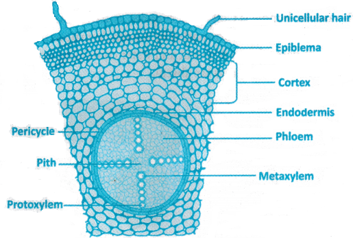
C) Stele:
It is the central part of the root and shows pericycle, vascular tissues, pith and conjunctive tissue.
i) Pericycle:
- Below the endodermis, a single layer of thin walled parenchymatous cells without intercellular spaces is present called the pericycle.
- These cells help in producing lateral roots and also become meristematic during secondary growth.
ii) Vascular tissues:
- Xylem and phloem strands are present on different radii so called radial vascular bundles.
- The xylem is in exarch type i.e.,protoxylem is towards the periphery and metaxylem is towards the centre.
- The xylem and the phloem are present in two to four patches so it is in Diarch to tetrarch condition.
iii) Conjunctive tissue:
- The parenchymatous tissue present between xylem and the phloem is called conjunctive tissue.
- It becomes meristematic during secondary growth.
D) Pith:
It is small, made up of parenchyma tissue or absent.
I. Multiple Choice Questions
Question 1.
Which of the following is not a component of stomatal apparatus?
1. Guard cell
2. Subsidiary cell
3. Trichome
4. Stomatal aperture
Answer:
3. Trichome
Question 2.
When xylem and phloem are jointly situated along the same radius in a vascular bundle it is called
1. Closed
2. Open
3. Radial
4. Conjoint
Answer:
4. Conjoint
Question 3.
A plant material which shows small pith, two to four xylem patches alternating with phloem.
1. Dicot root
2. Monocot root
3. Dicopt stem
4. Monocot stem
Answer:
1. Dicot root
Question 4.
The hypodermis is collenchymatous in
1. Monocot stem
2. Dicot stem
3. Dicot root
4. Both dicot stem and monocot root
Answer:
2. Dicot stem
Question 5.
Phloem parenchyma is absent in
1. Monocot stem
2. Dicot stem
3. Dicot root
4. Both dicot stem and monocot root
Answer:
1. Monocot stem
Question 6.
Casparian strips are made up of water impermeable substance ___________
1. Inulin
2. Suberin
3. Chitin
4. Cutin
Answer:
2. Suberin
Question 7.
In a dicot root conjunctive tissue is generally present between
1. Xylem and phloem
2. Xylem and pericycle
3. Phloem and pericycle
4. Endodermis and pericycle
Answer:
1. Xylem and phloem
Question 8.
Open vascular bundles are seen in
1. Dicot root
2. Monocot root
3. Dicot stem
4. Monocot stem
Answer:
3. Dicot stem
Question 9.
The elongated parenchyma cells arranged vertically and parallel to each other
1. Hypodermis in dicot stem
2. Palisade tissue in dicot leaf
3. Hypodermis in monocot stem
4. Bulliform cells of monocot leaf
Answer:
2. Palisade tissue in dicot leaf
Question 10.
Water containing cavities in the vascular bundles is a characteristic of
1. Dicot leaf
2. Dorsiventral leaf
3. Dicot stem
4. Monocot stem
Answer:
4. Monocot stem
II. Fill in the Blanks
Question 1.
The special cells that surround the guard cells in the stomatal apparatus are ___________
Answer:
Subsidiary cells
Question 2.
Simple tissues like parenchyma, collenchyma and sclerenchyma are largely present in the ___________ tissue system.
Answer:
Ground
Question 3.
The tissue present between xylem and phloem in a dicot stem is ___________.
Answer:
Cambium
Question 4.
In the anatomy of a dicot or monocot root all the tissues on the inner side of the endodermis such as pericycle, vascular bundles and pith constitute the ___________.
Answer:
Stele
Question 5.
The innermost layer of cortex is ___________
Answer:
Endodermis
Question 6.
The tissue between upper and lower epidermis in a leaf is ___________.
Answer:
Mesophyll
Question 7.
The large colourless cells in the adaxial surface of monocot leaf are ___________.
Answer:
Bulliform cells
Question 8.
The conjoint vascular bundles usually have the phloem located only on the ___________ side of Xylem.
Answer:
Outer
Question 9.
Mechanical strength to the young dicot stem is provided by hypodermis which is made up of ___________ cells.
Answer:
Collenchymatous
Question 10.
The cells of ___________ in dicot stem are rich in starch grains, hence this layer is also referred to as starch sheath.
Answer:
Endodermis
III One Word Answer Questions
Question 1.
To which tissue system does the roothair and trichomes belong?
Answer:
Epidermal tissue system
Question 2.
What name is given to the vascular bundles of roots where the xylem and phloem are arranged in an alternate manner along different radii?
Answer:
Radial vascular bundles / Alternate
Question 3.
What is the region between epidermis and stele in a dicot stem called?
Answer:
Cortex
Question 4.
Which cells of a grass leaf help in curling to minimise water loss?
Answer:
Bulliform cells
Question 5.
What do you call the loosely arranged tissue situated below the palisade in a dicot leaf?
Answer:
Spongy parenchyma
Question 6.
In which vegetative part (among stem, root and leaf) of a plant body cuticle is absent?
Answer:
Roots
Question 7.
With respect to the presence of cambium, what is the name given to the vascular bundle in a dicot stem?
Answer:
Open vascular bundle
Question 8.
Which is the part in the dicot stem with a large number of rounded, parenchymatous cells with intercellular spaces in the stele?
Answer:
Pith or medulla
Question 9.
What is the shape of sclerenchymatous patches of pericycle in a dicot stem?
Answer:
Semilunar
Question 10.
In a dorsiventral leaf, which epidermis bears more stomata?
Answer:
Lower (Abaxial) epidermis