AP Inter 1st Year Zoology 8th Lesson Neural Control and Coordination Questions and Answers
IV. Very Short Answer Questions
Question 1.
Distinguish between afferent and efferent nerve fibres.
Answer:
The nerve fibres of the PNS are of two types.
a) Afferent fibres (sensory): The afferent nerve fibres transmit impulses from tissues/ organs to the CNS.
b) Efferent fibres (Motor): The efferent fibres transmit regulatory impulses from CNS to concerned tissues/ organs.
Question 2.
Where do we find myelinated and non-myelinated nerve fibres ?
Answer:
- Myelinated nerve fibres are found in spinal and cranial nerve. Spinal/cranial nerves form SNS.
- Unmyelinated nerve fibre are commonly found in autonomous and somatic neural systems.
Question 3.
Distinguish between unipolar and bipolar neurons.
Answer:
- Unipolar: Cell body with one axon, found usually in embryonic stage.
- Bipolar: One axon and one dendrite eg: retina of eye and olfactory membrane in nose.
Question 4.
Name the three cranial meninges covering the human brain.
Answer:
The skull of the brain is covered by cranial meninges consisting of an outer layer called Duramater (fibrous), a very thin middle layer called arachnoid and an inner layer which is in contact with the brain tissue called piamater (vascular).
Question 5.
What is corpus callosum?
Answer:
- Corpus callosum is a transverse band internal connection between two cerebral hemispheres beneath the cerebral cortex.
- It is made up of a wide, flat bundle of myelinated fibres. It brings co-ordination between two cerebral hemispheres.
Question 6.
What is a synapse?
Answer:
Synapse: It is formed by the membranes of a pre-synaptic and a post-synaptic neuron that may or may not be separated by a gap known as the synaptic cleft. There are two types of synapses, namely chemical synapses and electrical synapses.
Question 7.
Distinguish between the grey matter and the white matter of the central neural system.
Answer:
- Grey matter, primarily composed of neuronal cell bodies and dendrites.
- White matter, composed mainly of myelinated axons.
Question 8.
What are the corpora quadrigemina?
Answer:
- Dorsal portion (posterior) of the mid brain consists mainly of four round swellings (lobes) called as corpora quadrigemina.
- One pair is superior colliculi (visual activity) and one pair inferior colliculi (auditory activity) both are a part of corpora quadrigemina.
V. Short Answer Questions
Question 1.
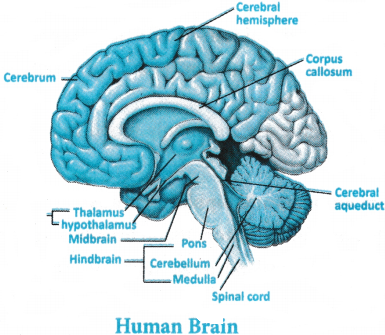
Draw a labeled diagram of the human brain.
Answer:
Question 2.
Compare the following.
a) Central neural system (CNS) and Peripheral neural system (PNS).
b) Resting potential and Action potential.
Answer:
a) Central neural system (CNS) and Peripheral neural system (PNS)
Central neural system (CNS):
- The spinal cord is protected by the vertebral column, whereas the brain is protected by the skull.
- Consists of the spinal cord and the brain.
- Processes information and regulates the responses to impulses.
Peripheral neural system (PNS):
- Nerves of PNS pass impulses to the CNS and responses from the CNS to various structures of the body.
- It consists of the spinal nerves and the cranial nerves.
- It is divided into the autonomic nervous system and the somatic nervous system.
B) Resting potential and Action potential
Resting potential:
- When the neuron is at the resting phase, it is the potential difference across membrane.
- To maintain the resting potential, the sodium-potassium ATPase pump is activated, sending Na+ ions outside the neuron.
- Permeability of K+ ions is observed to be more by the plasma membrane of neurons.
- The exterior side of the neuron is positively charged, while the interior side is negatively charged.
Action potential:
- When the neuron is triggered, it is the potential difference across the membrane.
- It functions in a reverse pattern wherein the sodium-potassium ATPase pump sends Na+ ions to the neuron.
- The exterior side of the neuron is negatively charged, and the interior side of the neuron is positively charged.
- Permeability of Na+ ions is observed to be more by the plasma membrane of the neurons.
Question 3.
Give a brief account of the mechanism of synaptic transmission.
Answer:
Axon ending transfer nerve impulse to dendrite/cell body/axon of other neuron or to effector organ (e.g. muscle cell)
- A nerve is transmitted from one neuron to another through junctions called synapses.
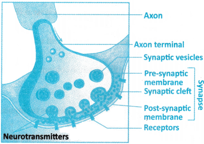
- A synapse if formed by membranes of a pre-synaptic neuron (previous neuron) and a post-Synaptic neuron (next neuron), which may or may not be separated by a gap called synaptic cleft.
Diagram showing axon terminal and synapse
There are two types of synapses, electrical synapses and chemical synapses.
1. Electrical synapses: The membrane of the pre and postsynaptic neurons are in very close proximity. Electrical current can flow directly from one neuron into other across these synapses.
- Transmission of an impulse in this case is very similar to impulse conduction along a single axon (Impulse directly transmitted from one neuron to another by making electric circuit).
- Impulse transmission across it is always faster than across a chemical synapse.
- Electrical synapses are rare in our system (but present in cardiac muscle fibre).
- Gap between two neurons is very less in these synapses and it is less energy consuming and there is no synaptic cleft. This synapse does not involve neuro transmitters.
2. Chemical synapse: The membrane of pre and post synaptic neurons is separated by a fluid space called synaptic cleft.
- Gap between two neurons is more, so synaptic cleft is present, impulse conduction is slow than electrical synapse and it is more energy consuming conduction. Most synapses in our neural system are of this kind.
- Chemicals or neurotransmitters are involved in the transmission of impulse at these synapses. The axon terminals contain vesicles filled with these neurotrans¬mitters.
- When an impulse/action potential arrives at axon terminal then voltage gated calcium channel open at terminal or synaptic knob and calcium ions enters in axon from extracellular fluid, this stimulates movement of synaptic vesicles towards pre synaptic membrane where they fuse with pre-synaptic membrane so, vesicles release their neurotransmitters (by exocytosis) in synaptic cleft.
- Released neurotransmitters bind their specific receptors, present on post-synaptic membrane. This binding opens ion channels on post synaptic membrane and allowing entry of ions which can generate a new potential in post-synaptic neuron
- New potential developed may be either excitatory or inhibitory depending on type of Neuro-transmitters.
Question 4.
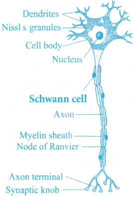
Describe the structure of a multipolar neuron with a labeled diagram.
Answer:
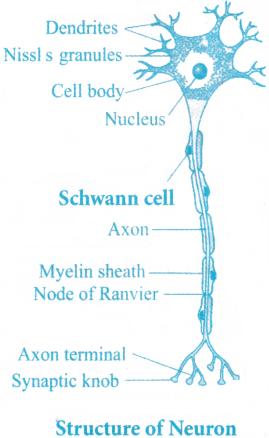
A neuron is a microscopic structure composed of three major parts, namely cell body, dendrites and axon.
Cell body:
- The cell body contains cytoplasm with typical cell organelles and certain granular bodes called Nissl’s granules (mass of ribosome’s, RER, these help in protein formation).
- It also contains neuro-fibrils and provides support to neuron.
Dendrites (short fibre):
- Which branch repeatedly and projects out from cell body, these also contain Nissl’s granules, and are called dendrites. These fibers transmit impulses towards the cell body.
- These are receiving end of information (response/stimuli) so known as afferent process.
Axon:
- It is a long fibre and distal end of which is branched.
- Each branch terminates as a bulb like structure called synaptic knob which possess synaptic vesicles containing neurotransmitters.
- The axon transmits nerve impulses away from cell body to a synapse (where two neurons meet) or to a neuro-muscular junction (Point of contact between neuron to muscle).
- Axon are delivering end of information (Stimulus/response) so known as efferent process.
- Axon part where, myelin sheath is absent is known as node of Ranvier but neurilemma is present (Layer of Schwann cell) at this place.
- In myelinated neuron, Schwann secretes myelin sheath which acts as electrical insulator.
- Based on the number of axon and dendrites, the neurons are divided into three types.
- Multipolar: One axon and two or more dendrites, found in cerebral cortex.
- Bipolar: One axon and one dendrite eg: retina of eye and olfactory membrane in nose.
- Unipolar: cell body with one axon, found usually in embryonic stage.
Pseudo unipolar: Present in dorsal root ganglia of spinal cord.
- On the basis of myelin sheath there are two types of axons, namely Myelinated and Non-Myelinated.
- The myelinated nerve fibers are enveloped with Schwann cells, which form myelin sheath around the axon.
- The gaps between two adjacent myelin sheaths are called Nodes of Ranvier.
- Myelinated nerve fibers are found in spinal and cranial nerve. Spinal/cranial nerves form SNS.
- Unmyelinated nerve fiber is enclosed by Schwann cell that does not form a myelin sheath around the axon, and is commonly found in autonomous and somatic neural systems.
Structure of Neuron
Question 5.
Differentiate between
a) Myelinated and Non-myelinated axons.
b) Dendrites and Axons.
a) Myelinated and Non-myelinated axons.
Answer:
On the basis of myelin sheath there are two types of axons, namely Myelinated and Non-Myelinated.
- The myelinated nerve fibres are enveloped with Schwann cells, which form myelin sheath around the axon.
- The gaps between two adjacent myelin sheaths are called Nodes of Ranvier.
- Myelinated nerve fibres are found in spinal and cranial nerve. Spinal/cranial nerves form SNS.
- The sheath acts as insulator, prevent energy loss in nerve transmission, these neurons show salutatory conduction.
- Unmyelinated nerve fibre is enclosed by Schwann cell that does not form a myelin sheath around the axon, and is commonly found in autonomous and somatic neural systems.
b) Dendrites and Axons.
Answer:
Dendrites: Dendrites are short processes, They are branched, Nissl’s granules are found in neuroplasm, They carry impulses towards the cell body of the neuron.
Axons: They are long processes, Axons may or may not be branched, Nissl’s granules are absent in neuroplasm, They carry impulses away from the cell body to the neuron.
Question 6.
Differentiate between
a) Thalamus and Hypothalamus.
b) Cerebrum and Cerebellum.
a) Thalamus and Hypothalamus.
Answer:
Thalamus: Consists of grey matter only and is located above the midbrain, It has the centre for sensations namely – cold, pain, heat. It does not secrete hormones.
Hypothalamus: Consists of white and grey matter and is located at the base of the thalamus, it has the centre for sensations namely – regulating body temperature, homeostasis, blood pressure. It secretes several hormones that control the activity of pituitary gland.
b) Cerebrum and Cerebellum.
Answer:
Cerebrum: It is a portion of the forebrain, The brain is majorly covered by the cerebrum, It is divided into two cerebral hemispheres, It is the centre for intelligence and memory.
Cerebellum: It is part of the hindbrain, The second largest part of the brain after the cerebrum, It is divided into three lobes, It is the centre for posture and body equilibrium.
Question 7.
Draw a labeled diagram of multipolar neuron.
Answer:
VI. Long Answer Questions
Question 1.
Give a brief account of the structure and functions of human brain with a neat labeled
diagram.
Answer:
- Brain is central information processing organ of our body, and acts as the command and control system.
- It controls the voluntary movements, balance of the body, functioning of vital involuntary organs (Ex: Lungs, Heart, Kidneys), thermoregulation, hunger and thirst, circadian rhythms (12 hours) of our body, and also control activities of several endocrine glands and human behaviour.
- It is also the site for processing of vision, hearing, speech, memory, intelligence, emotions and thoughts.
- The human brain is well protected by the skull.
- Inside the skull the brain is covered by cranial meninges consisting of an outer layer called Duramater (fibrous), a very thin middle layer called arachnoid and an inner layer which is in contact with the brain tissue called piamater (vascular).
- The brain can be divided into three major parts -Fore brain, mid brain and hind brain.
1. Fore brain: It consists of cerebrum, thalamus and hypothalamus.
Cerebrum:
- It forms major part of the human brain.
- A deep cleft (longitudinal fissure) divides cerebrum into longitudinally into two halves known as left and right cerebral hemispheres.
- Both hemispheres are connected by a tract of nerve fibres called Corpus callosum.
- The outer layer of cells which covers cerebral hemispheres is cerebral cortex, which is thrown (divided) into prominent (visible) folds known as gyri (outgrowth) and sulci (Depression).
- Cerebral cortex is outer part and contains grey matter, its greyish appearance is due to more neuronal cell bodies and non-myelinated neurons.
- Cerebral cortex contains rhotor areas to control voluntary activities and has association areas; these are neither clearly sensory nor motor in function but responsible for complex functions like inter sensory associations (detection of many senses at a time), memory and communication.
- Cerebral medulla constitutes the inner part of cerebral hemisphere. It has tracts of nerve fibers, covered with the myelin sheath.
- They give an opaque white appearance to this layer and hence are called as white matter.
- Cerebrum wraps (cover) around a structure called thalamus, which is major coordinating center for sensory and motor signaling.
- Another important part of the brain are Hypothalamus Lies at the base of thalamus. It contains a number of centers which controls body temperature, urge for eating (appetite) and drinking etc.
- It has several group of neurosecretory cells, which secrete hormones called hypothalamic hormones.
- Inner parts of cerebral hemispheres and a group of associated deep structures like amygdale, hippocampus etc. form complex structures called limbic lobe or limbic system. Along with hypothalamus, limbic system is involved in regulation of sexual behavior, expression of emotional reactions (Excitement, pleasure and happiness, rage or anger and fear) and motivation.
2. Mid brain:
- Located between thalamus/hypothalamus (both are part of diencephalon) of .fore brain on upper side and pons of mid brain on lower side.
- A canal called Aqueduct passes through the mid brain.
Dorsal portion (posterior) of the mid brain consists mainly of four round swellings (lobes) called as corpora quadrigemina.
3. Hind brain:
- It comprises of pons, cerebellum and medulla oblongata.
- Pons: Acts like a bridge and consists of tracts of fibres that interconnect different regions of the brain.
- Cerebellum: It has very convoluted (folded) surface in order to provide additional space for many more neurons.
Medulla oblongata:
- It connected to the spinal cord contains centers which controls respiration (Respiratory center), cardiovascular reflexes (cardiac center) and gastric secretions.
- Brain stem is made of 3 major regions they are mid brain, pons and medulla oblongata.
- Brain stem connects between the brain (cerebrum) and spinal cord.
Question 2.
Describe the mechanism of generation and conduction of nerve impulse through a nerve fibre with the help of diagrams.
Answer:
Diagram representation of Impulse conduction through an axon
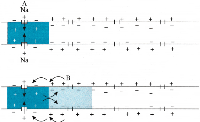
- Neurons are excitable cells because their membranes are in polarized state. Different types of ion channels are present on neural membrane.
- These ion channels are selectively permeable to different ions.
- When a neuron is not conducting any impulse.
- In resting state, the axonal membrane is comparatively more permeable to potassium ions (because K+ leak channels are more so it flows outward more) and nearly impermeable to Na+ ions (number is less, it moves inward less) and impermeable to negatively charged proteins present in the axoplasm (these remains inside of axon).
- The membrane is impermeable to negatively charged proteins present in the axoplasm. Consequently, the axoplasm inside the axon contains high concentration of K+ and negatively charged proteins and low concentration of Na+.
- The fluid outside the axon contains a low concentration of K+, a high concentration of Na+ and thus form a concentration gradient. These ionic gradients across the resting membrane are maintained by the active transport of ions by the sodium-potassium pump which transports 3Na+ outwards and 2K+ into cell.
- As a result, outer surface of axonal membrane possess a positive charge while its inner surface becomes negatively charged and this state is known as polarized-/resting state.
- When a stimulus is applied at site A on polarized membrane at site A becomes freely permeable to Na+. This leads to a rapid influx of Na+ followed by reversal of the polarity at site A outer surface of membrane becomes negatively charged and inner side becomes positively charges.
- The polarity of membrane at site A is thus reversed and hence depolarized.
- The electrical potential difference across plasma membrane at site A is now called as action potential, which is also termed as a nerve impulse.
- At sites immediately ahead the axon at site B, membrane has a positive charge on outer surface and a negative charge on its inner surface. As a result, a current flow on inner surface from site A to site B on outer surface current flows from site B to site A to complete circuit of current flow.
- So polarity at B site is reversed hence an action potential is generated at site B.
- Thus the impulse (action potential) generated at site A now arrives at site B. The sequence is repeated along length of axon and consequently impulse is conducted.
- The rise in stimulus induced permeability to Na+ is extremely short lived.
- It is quickly followed by a rise in permeability to K+.
- With in a fraction of a second K+ diffuses outside the membrane and it restores resting potential of membrane at site of excitation and it is known as repolarization and nerve fibre becomes ready for further stimulation.
I. Multiple Choice Questions
Question 1.
Which two systems jointly coordinate and integrate all the activities of the organs in human body ?
1. Neural system and Circulatory system
2. Endocrine system and Digestive system
3. Neural system and Endocrine system
4. Respiratory system and Neural system
Answer:
3. Neural system and Endocrine system
Question 2.
| Type of Neuron | Found in |
| a) Multipolar | i) Embryonic stage |
| b) Bipolar | ii) Cerebral cortex |
| c) Unipolar | iii) Retina of eye |
1. (a) i, (b) iii, (c) ii
2. (a) iii, (b) ii, (c) i
3. (a) ii, (b) iii, (c) i
4. (a) ii, (b) i, (c) iii
Answer:
3. (a) ii, (b) iii, (c) i
Question 3.
Sodium potassium pump transports,
1. Three K+ outwards for two Na+ into the cell.
2. Two Na+ outwards for three K+ into the cell.
3. Two K+ outwards for three Na+ into the cell.
4. Three Na+ outwards for two K+ into the cell.
Answer:
4. Three Na+ outwards for two K+ into the cell.
Question 4.
Depolarization of an axon occurs due to
1. Influx of Na+
2. Outflow of Na+
3. Influx of K+
4. Outflow of K+
Answer:
1. Influx of Na+
Question 5.
Which organ acts as “the command-and-control system of our body ?
1. Heart
2. Lungs
3. Brain
4. Kidney
Answer:
3. Brain
Question 6.
What is the correct sequence of meninges from inner to outer side ?
1. Arachnoid, Duramater, Piamater
2. Duramater, Arachnoid, Piamater
3. Piamater, Arachnoid, Duramater
4. Duramater, Piamater, Arachnoid
Answer:
3. Piamater, Arachnoid, Duramater
Question 7.
Which part of the human brain is the most developed ?
1. Cerebral cortex
2. Thalamus
3. Hypothalamus
4. Medulla
Answer:
1. Cerebral cortex
Question 8.
The limbic system is located in the
1. Cerebral hemispheres
2. Cerebellum
3. Midbrain
4. Medulla
Answer:
1. Cerebral hemispheres
Question 9.
What is the function of thalamus ?
1. To control body temperature.
2. To regulate hunger and thirst.
3. To act as a major coordinating centre for sensory and motor signaling.
4. To process vision and hearing.
Answer:
3. To act as a major coordinating centre for sensory and motor signaling.
Question 10.
What are the three major regions of the brain stem ?
1. Forebrain, hindbrain and cerebral cortex.
2. Forebrain, midbrain and spinal cord.
3. Cerebrum, midbrain and hindbrain.
4. Midbrain, pons and medulla.
Answer:
4. Midbrain, pons and medulla.
II. Fill in the blanks
Question 1.
____________ granules are present in the cell body of a neuron.
Answer:
Nissil’s
Question 2.
The chemicals present in the synaptic vesicles are ____________.
Answer:
Neurotransmitters
Question 3.
The gaps between two adjacent myelin sheaths are called ____________.
Answer:
Nodes of Ranvier
Question 4.
Neurons are excitable cells because their membranes are in a state.
Answer:
Polarised
Question 5.
The fluid filled space that separates the membranes of the pre-synaptic and post-synaptic neurons at a chemical synapse is ____________.
Answer:
Synaptic cleft
Question 6.
The cerebral hemispheres are connected by a tract of nerve fibres known as ____________.
Answer:
Corpus callosum
Question 7.
The canal that passes through the midbrain is called ____________.
Answer:
Cerebral aqueduct
III. One Word Answer Questions
Question 1.
What is the structural and functional unit of the neural system?
Answer:
Neuron.
Question 2.
Which fibres transmit the impulses towards the cell body in a neuron?
Answer:
Dendrites
Question 3.
Name the type of neurons with one axon and two or more dendrites.
Answer:
Multipolar neuron.
Question 4.
Which part of the eye consists of bipolar neurons?
Answer:
Retina.
Question 5.
Name the cells that form myelin sheath around the nerve fibres.
Answer:
Schwann cells.
Question 6.
Which type of synapses allow faster transmission of nerve impulses?
Answer:
Electrical synapses.
Question 7.
Which part of the forebrain controls body temperature, urge for eating and drinking?
Answer:
Hypothalamus.