AP Inter 1st Year Zoology 7th Lesson Locomotion and Movement Questions and Answers
IV. Very Short Answer Questions
Question 1.
Write the differences between actin and myosin.
Answer:
Actin:
- Actin is thin contractile protein.
- It is present in thin filaments of a myofibril.
- Each actin filament is made of two ‘F’ actin molecules.
Myosin:
- Myosin is a thick contractile protein.
- Myosin is present as a thick filament in a dark band of a myofibril.
- Each myosin is made up of monomeric proteins called meromyosins.
Question 2.
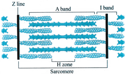
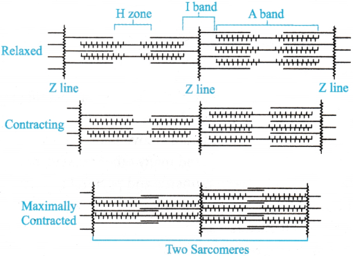
Draw the diagram of a sarcomere of skeletal muscle showing different regions.
Answer:
Question 3.
Distinguish between red muscle fibres and white muscle fibres.
Answer:
| Red muscle fibre | White muscle fibre |
| More myoglobin | Less myoglobin |
| Slow contraction | Fast contraction |
| More mitochondria | Less mitochondria |
| Aerobic respiration | Anaerobic respiration |
| Sarcoplasmic Reticulum is poorly developed. | Sarcoplasmic Reticulum is more developed. |
| Slow but longer time contraction. | Fast but shortlasting contraction. |
| Eg: Extensor muscles of back, Flight muscles of kite. | Eg: Muscles of eyeballs, flight muscles of sparrow. |
Question 4.
Name the type of joint between the following.
a) Atlas / Axis
b) Carpal/Metacarpal of the human thumb
Answer:
a) Joint between atlas and axis is pivot joint.
b) Joint between carpal and metacarpal of the human thumb is saddle joint.
Question 5.
Name the type of joint between
a) Cranial bones
b) Phalanges
Answer:
- Joint between cranial bones is sutures (Fibrous joint).
- The joints between phalanges are hinge joints.
Question 6.
Human skull is described as dicondylic skull. Give the reason.
Answer:
Human skull contains one occipital condyle on each side of the foramen magnum. Hence it is called dicondylic skull.
Question 7.
Name the ear ossicles in human beings.
Answer:
Ear ossicles are three in number in the middle car of human beings.
- Malleus-hammer shaped outer bone. It is derived from articular bone of lower jaw of earlier vertebrates.
- Incus-anvil shaped middle bone. It is derived from quadrate bone of upper jaw.
- Stapes- stirrup shaped inner bone.
It is derived from hyomandibula of lower vertebrates.
V. Short Answer Questions
Question 1.
Write a short note on sliding filament theory of muscle contraction.
Answer:
The process of muscle contraction involves various steps.
a) Excitation of muscle: When a signal from CNS reaches to the neuro muscular j unction, a neurotransmitter ‘acetyl-choline’ is released. Acetyl choline generates action potential in sarcolemmAnswer: The action potential passes to T-tubules then to terminal cisternae to release Ca++ ions into sarcoplasm.
b) Formation of cross bridges: The Ca++ ions bind to Tn-C of troponin. As a result, troponin and tropomyosin move away from active sites on actin filaments. Then the head of myosin by using energy from hydrolysis of ATP go and bind to active sites on actin filaments and form cross bridges.
c) Power stroke: The cross bridges pull the actin filaments over thick myosin filaments towards the center of A band, it is called power stroke. Thus, the length of sarcomere decreases.
d) Recovery stroke: The myosin goes back to its relaxed state and releases ADP. A new ATP molecule binds to the head of myosin and the cross bridge is broken; it is called recovery stroke.
e) Relaxation of muscle: When the motor impulses from CNS stops Ca++ ions are pumped back to sarcoplasmic reticulum. Active sites are covered by troponin, tropomyosin complex. Z-lines of sarcomere reaches to original position.
Question 2.
Describe the structure of a skeletal muscle.
Answer:
Each skeletal muscle in our body is made up of a number of muscle bundles or fascicles. Each fascicle is covered by a connective tissue sheath called perimysium.
- All the fascicles of a striated muscle are connected by a common collagenous connective tissue layer known as fascia. Each fascicle is consisting of several number of cylindrical muscle fibres. Each muscle fibre is covered by a thin connective tissue sheath called endomysium.
- Each muscle fibre or muscle cell shows sarcolemma, sarcoplasm, many peripheral nuclei (structural syncytium), sarcoplasmic reticulum, sarcosomes, reserve energy molecules, myoglobin, myofibrils etc.
- Each myofibril is made of thin actin filaments and thick myosin filaments. Each myofibril shows alternate arrangement of dark and light bands. So, the skeletal muscle is striated.
- The unit of muscle contraction is sarcomere. It is a part of myofibril present in between two successive ‘Z’ lines. It consists of a middle complete A band and two half I-bands on either side of it.
- In a skeletal muscle, triad systems are present at A-T junctions of myofibril.
Question 3.
Write short notes on contractile proteins.
Answer:
In a myofibril, there are two kinds of filaments namely.
a) Thin filament: It is made of three kinds of proteins.
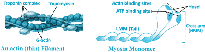
i) Actin: It occurs in the form of two filamentous molecules (two F-actins) that are coiled helically around each other. Each ‘F-actin is made up of several monomers of ‘G’ actin molecules.
ii) Troponin: It is made up of three polypeptide units named as Tn-I, Tn-C and Tn-T. Tn-T binds to tropomyosin. T-I inhibits myosin n binding site on actin. T1-C binds to Ca++ ions. When Ca++ ions hind to T-C active sites open so that myosin heads bind to active sites and form actomyosin complex.
iii) Tropomyosin: It runs close to ‘F’ actin molecules all along the length of actin. Both troponin and tropomyosin are often called as regulatory proteins because they are going to mask or unmask active sites.
b) Thick filament:
- Each thick filament is made up of myosin proteins.
- Each thick filament contains 200-300 molecules of myosin.
- Myosin: It is considered as a motor protein, as it convert chemical energy to mechanical energy. Each myosin is made up of several monomeric proteins called meromyosin.
- Each meromyosin has a globular head, short arm or neck and tail.
- Head is made up of heavy meromyosin (HMM) and the tail is made up of light meromyosin (LMM).
- Head has two binding sites one for ATP and other to bind with active site.
- The heads of myosin molecules are directed towards Z-membrane in the relaxed state. Myosin heads form cross bridges during contraction.
- Short arm serves as a flexible link between head and tail regions.
- The tails of myosin molecules are directed towards M – line of H-zone.
- The contractile proteins of thick and thin filaments involve in muscle contraction.
Question 4.
How do you distinguish between a skeletal muscle and a cardiac muscle?
Answer:
The differences between a skeletal muscle and a cardiac muscle are
| Skeletal Muscle | Cardiac Muscle |
| Linked to the primary bones. | Located in the walls of the heart. |
| Voluntary in nature. | Involuntary in nature. |
| Peripherally located nucleus. | Centrally located nucleus. |
| The muscle fibres of the skeletal muscles are unbranched. | The muscle fibres of the cardiac muscles are branched. |
| Intercalated discs are not found in the Muscle fibres of the skeletal muscles. | Intercalated discs are found in the muscle fibres of the cardiac muscles. |
| Brings about the locomotory actions of the body and maintains body posture. | Responsible for the movement/motion of the heart. |
Question 5.
Write short notes on the ribs of human beings.
Answer:
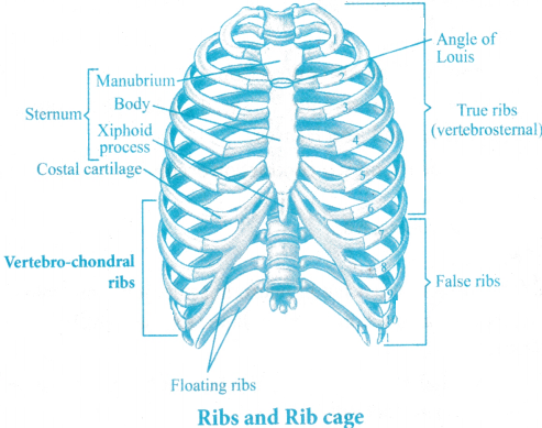
- Twelve pairs of ribs are present in the human. Ribs are thin flat curved bones, present in chest region and form the rib cage by connecting dorsally to vertebral column and ventrally to sternum.
- Rib cage gives protection to vital organs of the body. Ribs of man are called bicephalic as they have two heads.
- Ribs are categorized into true ribs and false ribs.
True ribs (vertebro-sternal ribs):
- First seven pairs are true ribs as they are connected to thoracic vertebrae on dorsal side and the sternum on ventral side.
- These ribs join to sternum by costal cartilages (hyaline cartilage).
False ribs:
- The remaining pairs of ribs are called false ribs. 8th, 9th and 10th pairs do not articulate directly with sternum but are connected to cartilagenous parts of seventh rib. Hence they are called vertebro-chondral ribs.
- The last two pairs (11th and 12th) are not connected to sternum or to anterior ribs, so they are called floating ribs.
Question 6.
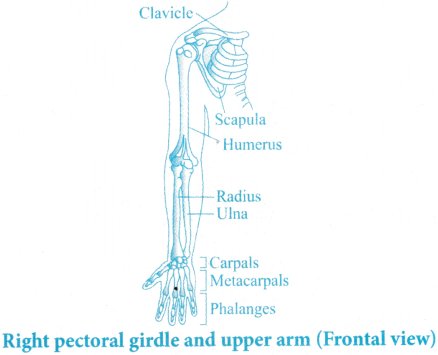
Draw a neat labelled diagram of the forelimb of man.
Answer:
Question 7.
Give an account of synovial joints.
Answer:
- Synovial joints are characterized by the presence of a fluid filled synovial cavity between the articulating surfaces of the two bones.
- Such arrangement allows considerable movement.
- These joints help in locomotion and many other movements.
- Ball and socket joint (between humerus and pectoral girdle).
- Hinge joint (knee j oint).
- Pivot joint (between atlas and axis).
- Gliding joint (between the carpals) and
- Saddle joint (between carpal and metacarpal of thumb).
VI. Long Answer Questions
Question 1.
Explain the mechanism of muscle contraction.
Answer:
The mechanism of muscle contraction was well explained by Sliding Filament Theory proposed by Hugh Huxley and Jean Hansen.
- According to this theory, muscle contraction occurs because of sliding of thin filaments over thick filaments.
Mechanism of muscle contraction:
Excitation of Muscle:
- A signal for contraction reaches to neuromuscular junction from the CNS.
- At the neuromuscular junction acetyl choline is released which generates action potential in sarcolemmAnswer:
- The action potential travels through T-tubule of the triad system and the cisternae release cations into sarcoplasm.
Stages in cross bridge formation, rotation of head and breaking of cross bridge
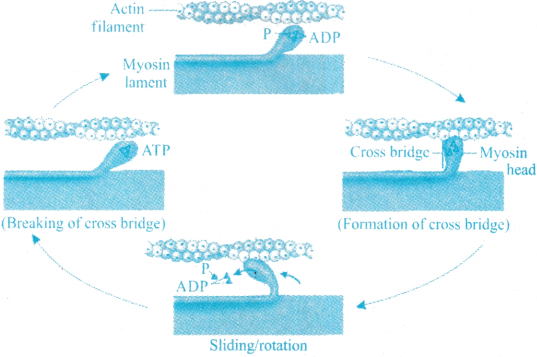
Sliding filament theory of muscle contraction
Formation of cross bridges:
- As the level of Ca++ ions increase, some Ca++ ions bind to Tn-C sub unit of troponin.
- Attachment of Cations to Tn-C makes troponin and tropomyosin to move away from active site.
- Thus, active sites are exposed on actin filaments Myosin head now binds to exposed active site by using the energy obtained by hydrolysis of ATP.
- Binding of myosin with active site on actin filaments results in formation of actomyosin complex.
- The head of myosin that fits into active site acts like a cross bridge (cross arm).
Power stroke:
- When myosin heads hydrolysis ATP into ADP and P, the conformation of the myosin is changed to an active state so that it can perform power stroke. The bending of cross arms towards H-zone is called power stroke.
- The actin filaments now slide over the myosin filaments. As a result, actin filaments are pulled towards the center of A-band. Thus Z-lines are also pulled inwards from both sides. Hence the sarcomere is subjected for contraction.
- During this process the length of sarcomere decreases, but the length of A-band remains constant, whereas the length of H-zone decreases. The muscle shows the effect of contraction now.
Recovery Stroke: Myosin goes back to relaxed state by releasing ADP.A new ATP molecule binds to head of myosin and cross bridges detach from active site.
Relaxation of Muscle: Motor impulses from CNS stops, rendering the cations to re-enter into sarcoplasmic cisternae of triad system. Active sites are now masked by the regulatory proteins (troponin and tropomyosin complex). Hence head of myosin cannot bind to active site on actin filaments. Z-membrane also reaches to original position hence the muscle relaxes.
I. Multiple Choice Questions
Question 1.
Why is the skeletal muscle fibre considered a syncytium?
1. It has multiple nuclei in the sarcoplasm.
2. It is surrounded by a single membrane.
3. It lacks a cytoskeleton.
4. It has a single nucleus.
Answer:
1. It has multiple nuclei in the sarcoplasm.
Question 2.
The filamentous actin (‘F actin) is made up of
1. Tropomyosin polymers
2. G actin (Globular actin) monomers
3. Myosin filaments
4. Troponin complexes
Answer:
2. G actin (Globular actin) monomers
Question 3.
Meromyosin is composed of
1. Only heavy meromyosin (HMM).
2. Heavy meromyosin (HMM) and light meromyosin (LMM).
3. Tropomyosin and troponin.
4. ‘F’ actin and ‘G’ actin.
Answer:
2. Heavy meromyosin (HMM) and light meromyosin (LMM).
Question 4.
Which neurotransmitter is released at the neuromuscular junction?
1. Dopamine
2. Acetylcholine
3. Serotonin
4. Norepinephrine
Answer:
2. Acetylcholine
Question 5.
ATP hydrolysis provides energy for
1. Release of neurotransmitters at the neuromuscular junction.
2. Binding of calcium to troponin.
3. Relaxation of the sarcolemma.
4. Cross-bridge formation.
Answer:
4. Cross-bridge formation.
Question 6.
Muscle fatigue is caused by
1. Excess caletum ions in the sarcoplasm.
2. Accumulation of lactic acid.
3. Increased oxygen levels in red fibres.
4. Depletion of myoglobin in white fibres.
Answer:
2. Accumulation of lactic acid.
Question 7.
Red muscle fibres are rich in
1. Sarcoplasmic reticulum and glycogen
2. Lactic acid and ATP
3. Myoglobin and mitochondria
4. Troponin and tropomyosin
Answer:
3. Myoglobin and mitochondria
Question 8.
The hyoid bone is located at the
1. Front part of the skull
2. Base of the buccal cavity
3. Superior region of the vertebral column
4. Nasal cavity
Answer:
2. Base of the buccal cavity
Question 9.
Identify the A, B, C, D parts from the figure given below.
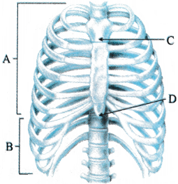
1. A-true ribs B-sternum C-false ribs D-vertebral column
2. A-true ribs B-false ribs C-D-vertebral column
3. A-false ribs B-vertebral column C-true ribs D-sternum
4. A-true ribs B-sternum C-vertebral column D-false ribs
Answer:
2. A-true ribs B-false ribs C-D-vertebral column
Question 10.
Each half of the human pelvic girdle is formed by the fusion of
1. Ilium, ischium, pubis
2. Ilium, femur, tibia
3. Pubis. Femur, ischium
4. Pubis, ilium, radius
Answer:
1. Ilium, ischium, pubis
Question 11.
The disorder caused by the accumulation of uric acid crystals in the joints
1. Arthritis
2. Osteoporosis
3. Gout
4. Muscular dystrophy
Answer:
3. Gout
Question 12.
Myasthenia gravis affects the
1. Cartilages
2. Joints
3. Neuromuscular junctions
4. Bone marrow
Answer:
3. Neuromuscular junctions
Question 13.
Match Column-I with Column-II
| Column-I | Column-II |
| A) Smooth muscle | i) Myoglobin |
| B) Tropomyosin | ii) Thin filament |
| C) Red muscle | iii) Sutures |
| D) Skull | iv) Involuntary |
Choose the correct option
1. A-i, B-iii, C-iv, D-ii
2. A-iv, B-i, C-ii, D-iii
3. A-iii, B-ii, C-i, D-iv
4. A-iv, B-ii, C-i, D-iii
Answer:
4. A-iv, B-ii, C-i, D-iii
II. Fill in the Blanks
Question 1.
The store house of calcium ions in a muscle fibre ___________.
Answer:
Sarcoplasmic reticulum
Question 2.
The functional unit of muscle contraction is ___________.
Answer:
Sarcomere
Question 3.
The Junction between a motor neuron and the sarcolemma of the muscle fibre is called ___________.
Answer:
Neuromuscular junction
Question 4.
The red coloured pigment that stores oxygen in the muscle is called ___________.
Answer:
Myoglobin
Question 5.
The number of cervical vertebrae in the human body ___________.
Answer:
Seven (7)
Question 6.
The two filaments that run close to the F’ actins in a myofibril are made up of ___________ protein.
Answer:
Tropomyosin
Question 7.
The flat bone located on the ventral midline of human thorax is ___________.
Answer:
Sternum
Question 8.
The longest bone in the human body is ___________.
Answer:
Femur
III. One Word Answer Questions
Question 1.
Which protein forms the thick filaments in a muscle fibre?
Answer:
Myosin.
Question 2.
What type of joint is found between femur and acetabulum?
Answer:
Ball and Socket joint.
Question 3.
What disorder is characterized by inflammation of joints?
Answer:
Arthritis.
Question 4.
What is the first vertebra of vertebral column called?
Answer:
Atlas.
Question 5.
Which bone is commonly known as collar bone in man?
Answer:
Clavicle.
Question 6.
Which skeletal disorder is characterized by decreased bone mass?
Answer:
Osteoporosis.
Question 7.
How many pairs of ribs are present in the human body?
Answer:
12.
Question 8.
What is the cup-shaped bone covering the knee joint?
Answer:
Patella.