AP Inter 1st Year Zoology 5th Lesson Body Fluids and Circulation Questions and Answers
IV. Very Short Answer Questions
Question 1.
What are the antibodies of ABO blood grouping ? Where are they present ?
Answer:
- Blood type A persons have anti- B antibodies, blood type B persons have anti- A antibodies, the blood type AB persons have no antibodies and blood type O persons have both anti- A and anti- B antibodies in plasma.
- ABO blood grouping system has antibodies A and B in blood plasma.
Question 2.
What is erythroblastosis foetalis ?
Answer:
The Rh antibodies of the sensitized Rh negative mother react with Rh antigen of positive foetus leading to destruction of foetal RBC. This disorder is called erythroblastosis foetals.lt occurs in an Rh+ve foetus, whose father is Rh+ve and the mother is Rh-ve.
Question 3.
Sino-atrial node is called the pacemaker of our heart. Why ?
Answer:
Sino-atrial node (SAN) is called the ‘pacemaker’ because it has the ability to generate action potentials without any external stimuli. It initiates heartbeat. It lies in the right upper part of right atrium.
Question 4.
Name the valves that guard the atrioventricular apertures in man.
Answer:
Left atrioventricular aperture is guarded by ‘bicuspid valve’ (mitral valve).Right atrioventricular aperture is guarded by tricuspid valve.
Question 5.
Name the heart sounds. When are they produced ?
Answer:
- First heart sound is called “Lub”. It is produced in ventricular systole due to closure of the AV valves.
- Second heart sound is called “Dub”. It is produced in cardiac diastole due to closure of semilunar valves.
Question 6.
Define cardiac cycle and cardiac output.
Answer:
Cardiac cycle:
The cardiac events that occur from the beginning of one heart beat to the beginning of the next are collectively called cardiac cycle.
Cardiac output:
The volume of blood pumped out by the heart from each ventricle per minute is called cardiac output.
Cardiac output = Stroke volume × No. of beats per minute.
70 ml × 72 beats/minute = 5040 ml/min. or approximately 5 liters.
Question 7.
What is meant by double circulation ? What is its significance ?
Answer:
Double circulation: It is the pattern of blood circulation in which blood passes through the heart twice in a complete circuit of blood. It includes pulmonary and systemic circulations.
Significance: The systemic circulation provides nutrients, O2 and other essential substances to the tissues and collects CO2 and other harmful substances away, for their elimination.
The pulmonary circulation eliminates CO2 into alveolar air and collects O2 from the alveolar air. Mixing of the oxygenated and deoxygenated types of blood is prevented.
Question 8.
What is electrocardiogram and what are the normal components of it ?
Answer:
Electrocardiogram is the technique by which the electrical activities of the heart are studied. The normal components of ECG include
- Waves (P.Q.R.S and T waves)
- Intervals (P-R; Q-T; R-R intervals)
- Segments (S-T segment)
- Complexes (QRS complex)
Question 9.
Name the three layers of blood vessels.
Answer:
Basically, each artery and vein consists of three layers: an inner lining of squamous endothelium, the tunica intima, a middle layer of smooth muscle and elastic fibers, the tunica media, and an external layer of fibrous connective tissue with collagen fibers, the tunica externAnswer:The tunica media is comparatively thin in the veins.
V. Short Answer Questions
Question 1.
Name the components of the formed elements in the blood and mention one major function of each of them.
Answer:
Formed Elements:
Erythrocytes, leucocytes and platelets are collectively called formed elements and they constitute nearly 45% of the blood.
Blood = Plasma + RBC + WBC + Platelets.
(a) Erythrocytes or Red blood cells (RBC):
- These are the most abundant of all the cells in the blood.
- A healthy adult man has, on an average, 5 million to 5.5 millions of RBCs mm’3 of blood.
- RBCs are formed in the red bone marrow in the adults.
- RBCs are devoid of nucleus in most of the mammals and are biconcave in shape
- They have a red coloured, iron containing complex protein called haemoglobin, hence the colour and name of these cells.
- A healthy individual has 12-16 grams of haemoglobin in every 100 ml of blood.
- These molecules play a significant role in transport of respiratory gases.
- RBCs have an average life span of 120 days after which they are destroyed in the spleen (graveyard of RBCs).
(b) Leucocytes:
- These are also known as white blood cells (WBC) as they are colourless due to the lack of haemoglobin.
- They are nucleated and are relatively lesser in number which averages 6000 – 8000 mm3 of blood.
- Leucocytes are generally short lived. We have two main categories of WBCs – Granulocytes and Agranulocytes.
- Neutrophils, Eosinophils and basophils are different types of granulocytes, while lymphocytes and monocytes are the Agranulocytes.
- Neutrophils are the most abundant cells (60-65 %) of the total WBCs and basophils are the least (0.5-1%) among them.
- Neutrophils and monocytes (6-8%) are phagocytic cells which destroy foreign organisms entering the body.
- Basophils secrete histamine, serotonin, heparin etc. and are involved in inflammatory reactions.
- Eosinophils (2-3%) resist infections and are also associated with allergic reactions.
- Lymphocytes (20-25%) are the two major types – ‘B’ and T forms. Both B and T lymphocytes are responsible for immune responses of the body.
(c) Platelets:
- These are also called Thrombocytes are cell fragments produced from megakaryocytes (special cells in the bone marrow).
- Blood normally contain 1,50,000 – 3,50,000 platelets mm-3.
- Platelets can release a variety of substances most of which are involved in the coagulation or clotting of blood.
- A reduction in their number can lead to clotting disorders which will lead to excessive loss of blood from the body.
Question 2.
Explain the mechanism of clotting of blood.
Answer:
- Blood exhibits coagulation or clotting in response to an injury or traumAnswer:This is a mechanism to prevent excessive loss of blood from the body.
- A dark reddish brown scum formed at the site of a cut or an injury over a period of time.
- It is a clot or coagulum formed mainly of a network of threads called fibrins in which dead and damaged formed elements of blood are trapped.
- Fibrins are formed by the conversion of inactive fibrinogens in the plasma by the enzyme thrombin.
- Thrombins, in turn are formed from another inactive substance present in the plasma called prothrombin.
- An enzyme complex, thrombokinase, is required for the above reaction.
- This complex is formed by a series of linked enzymic reactions (cascade process), involving a number of factors present in the plasma in an inactive state.
- An injury or a trauma stimulates the platelets in the blood to release certain factors which activate the mechanism of coagulation.
- Certain factors released by the tissues at the site of injury also can initiate coagulation.
- Calcium ions play a very important role in clotting.
Question 3.
Write short notes on ABO blood grouping.
Answer:
ABO grouping:
- It is based on the presence or absence of two surface antigens (chemicals that can induce immune response) on the RBCs namely A and B.
- Similarly, the plasma of different individuals contains two natural antibodies (proteins produced in response to antigens).
- The distribution of antigens and antibodies in the four groups of blood A, B, AB and O.
Blood Groups and Donor Compatibility
| Blood Group | Antigens on RBCs | Antibodies in Plasma | Donor’s Group |
| A | A | Anti-B | A, O |
| B | B | Anti-A | B, O |
| AB | A, B | nil | AB, A, B, O |
| O | nil | Anit-A, B | O |
- During blood transfusion, any blood cannot be used; the blood of a donor has to be carefully matched with the blood of a recipient before any blood transfusion to avoid severe problems of clumping (destruction of RBC).
- It is evident that group ‘O’ blood can be donated to person with any other blood group and hence ‘O’ group individuals are called ‘Universal donors’.
- Persons with ‘AB’ group can accept blood from persons with AB as well as the other groups of blood. Therefore, such persons are called ‘Universal recipients’.
Question 4.
Describe erythroblastosis foetalis.
Answer:
- A special case of Rh incompatibility (mismatching) has been observed between the Rh-ve blood of a pregnant mother with Rh+ve blood of the foetus.
- Rh antigens of the foetus do not get exposed to the Rh-ve blood of the mother in the first pregnancy as the two bloods are well separated by the placenta.
- However, during the delivery of the first child, there is a possibility of exposure of the maternal blood to small amounts of the Rh+ve blood from the foetus.
- In such cases, the mother starts preparing antibodies against Rh antigen in her blood.
- In case of her subsequent pregnancies, the Rh antibodies from the mother (Rh-ve) can leak into the blood of the foetus (Rh+ve) and destroy the foetal RBCs.
- This could be fatal to the foetus or could cause severe anaemia and Jaundice to the baby. This condition is called erythroblastosis foetalis.
- This can be avoided by administering anti-Rh antibodies to the mother immediately after the delivery of the first child.
Question 5.
Write a short note on lymph.
Answer:
LYMPH (Tissue fluid):
- As the blood passes through the capillaries in tissues, some water along with many small water soluble substances move out into the spaces between the cells of tissues leaving the larger proteins and most of the formed elements in the blood vessels.
- This fluid released out is called the interstitial fluid or tissue fluid.
- It has the same mineral distribution as that in plasma.
- Exchange of nutrients, gases, etc., between the blood and the cells always occur through this fluid.
- An elaborate network of vessels called the lymphatic system collects this fluid and drains it back to the major veins.
- The fluid present in the lymphatic system is called the lymph. Lymph-is a colourless fluid containing specialized lymphocytes which are responsible for the immune responses of the body.
- Lymph is also an important carrier for nutrients, hormones, etc.
- Fats are absorbed through lymph in the lacteals present in the intestinal villi.
Question 6.
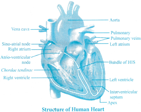
Draw a labelled diagram of the L.S of heart of man.
Answer:
Question 7.
Describe the atria of the heart of man.
Answer:
- Atria are thin walled blood ‘receiving chambers’ (upper chambers).
- The right one is larger than the left. The two atria are separated by thin inter-atrial septum. In the foetal heart, the atrial septum has a small pore called foramen ovale.
- In adults, fossa ovalis is present in the inter atrial septum. Fossa ovalis marks the position of the foetal foramen ovale. Right atrium receives deoxygenated blood from different parts of the body (except the lungs) through three caval veins.
- The openings of the precaval veins into the right atrium have no valves.
- The opening of the postcaval vein is guarded by the Eustachian valve. Eustachian valve directs the blood to the left atrium through the foramen ovale. In the foetal stage, but in the adult it becomes rudimentary and non-functional.
- Right atrium also receives blood from the myocardium (wall of the heart) through the coronary sinus. The opening of coronary sinus into the right atrium is guarded by the valve of Thebesius.
- Left atrium receives oxygenated blood from each lung through two pulmonary veins. The two left pulmonary veins open into the left atrium by a common aperture in some. The left and right atrioventricular apertures are guarded by bicuspid (mitral valve) and tricuspid valves respectively.
Question 8.
Describe the events in a cardiac cycle briefly.
Answer:
Cardiac Cycle
- There are four phases in a cardiac cycle.
Joint Diastole:
- To begin with, all the four chambers of heart are in a relaxed state, i.e., they are in joint diastole.
- As the tricuspid and bicuspid valves are open, blood from the pulmonary veins and vena cava flows into the left and the right ventricle respectively through the left and right atria.
- The semilunar valves are closed at this stage.
Atrial systole:
- The SAN now generates an action potential which stimulates both the atria to undergo a simultaneous contraction – the atrial systole.
- This increases the flow of blood into the ventricles by about 30 per cent.
- A specialised nodal tissue embedded in the lower corner of the right atrium, close to Atrio-ventricular septum, delays the spreading of impulse to heart approximately for about 0.1 seconds. This delay allows the atria to empty completely.
Ventricular systole:
- The action potential is conducted to the ventricular side by the AVN and AV bundle from where the bundle of His transmits it through the entire ventricular musculature.
- This causes the ventricular muscles contract, (ventricular systole), the atria undergoes relaxation (diastole), coinciding with the ventricular systole.
- Ventricular systole increases the ventricular pressure causing the closure of tricuspid and bicuspid valves due to attempted backflow of blood into the atria.
- As the ventricular pressure increases further, the semilunar valves guarding the pulmonary artery (right side) and the aorta (left side) are forced open, allowing the blood in the ventricles to flow through these vessels into the circulatory pathways.
Ventricular diastole:
- The ventricles now relax (ventricular diastole) and the ventricular pressure falls causing the closure of semilunar valves which prevents the backflow of blood into the ventricles.
- As the ventricular pressure declines further, the tricuspid and bicuspid valves are pushed open by the pressure in the atria exerted by the blood which was being emptied into them by the veins.
- The blood now once again moves freely to the ventricles. The ventricles and atria are now again in a relaxed (joint diastole) state, as earlier.
- Soon the SAN generates a new action potential and the events described above are repeated in that sequence and the process continues.
- This sequential event in the heart which is cyclically repeated is called the cardiac cycle and it consists of systole and diastole of both the atria and ventricles.
- The heart beats 72 times per minute, i.e., that many cardiac cycles are performed per minute. From this it could be deduced that the duration of a cardiac cycle is 0.8 seconds.
Stroke volume and Cardiac output:
- During a cardiac cycle, each ventricle pumps out approximately 70 mL of blood which is called the stroke volume.
- The stroke volume multiplied by the heart rate (no. of beats per min.) gives the cardiac output.
- Therefore, the cardiac output can be defined as the volume of blood pumped out by each ventricle per minute and averages 5000 mL or 5 liters in a healthy individual. During each cardiac cycle, the volume of blood pumped out by the right and left ventricle is same.
- Cardiac output = Stroke volume × Heart rate.
= 70 ml × 72/min = 5040 ml/min. - The body has the ability to alter the stroke volume as well as the heart rate and thereby the cardiac output. For example, the cardiac output of an athlete will be much higher than that of an ordinary man.
- During each cardiac cycle two prominent sounds are produced which can be easily heard through a stethoscope.
- The first heart sound (lub) is associated with the closure of the tricuspid and bicuspid valves whereas the second heart sound (dub) is associated with the closure of the semilunar valves.
Question 9.
Draw a standard ECG and explain the different segments in it.
Answer:
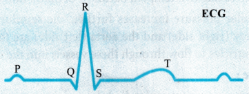
- ECG is a graphical representation of the electrical activity of the heart during a cardiac cycle.
- To obtain a standard ECG a patient is connected to the machine with three electrical leads (one to each wrist and to the left ankle) that continuously monitor the heart activity.
- For a detailed evaluation of the heart’s function, multiple leads are attached to the chest region. Here, we will talk only about a standard ECG.
- Each peak in the ECG is identified with a letter from P to T that corresponds to a specific electrical activity of the heart.
- The P-wave represents the electrical excitation (or depolarization) of the atria, which leads to the contraction of both the atria.
- The QRS complex represents the depolarization of the ventricles, which initiates the ventricular contraction.
- The contraction starts shortly after and marks the beginning of the systole.
- The T-wave represents the return of the ventricles from excited to normal state (repolarization). The end of the T-wave marks the end of systole.
Since the ECGs obtained from different individuals have roughly the same shape for a given lead configuration, any deviation from this shape indicates a possible abnormality or disease. Hence, it is of a great clinical significance.
VI. Long Answer Questions
Question 1.
Describe the structure of the human heart with the help of a neat labelled diagram.
Answer:
Heart, the mesodermally derived organ, is situated in the thoracic cavity, in between the two lungs, slightly tilted to the left. It has the size of a clenched fist.
- It is protected by a double walled membranous bag, pericardium, enclosing the pericardial fluid.
- Our heart has four chambers, two relatively small upper chambers called atria and two larger lower chambers called ventricles.
- A thin, muscular wall called the inter-atrial septum separates the right and the left atria, whereas a thick-walled, the inter-ventricular septum, separates the left and the right ventricles.
- The atrium and the ventricle of the same side are also separated by a thick fibrous tissue called the atrio-ventricular septum.
- However, each of these septa is provided with an opening through which the two chambers of the same side are connected.
- The opening between the right atrium and the right ventricle is guarded by a valve formed of three muscular flaps or cusps, the tricuspid valve, whereas a bicuspid or mitral valve guards the opening between the left atrium and the left ventricle.
- The openings of the right and the left ventricles into the pulmonary artery and the aorta respectively are provided with the semilunar valves.
- The valves in the heart allow the flow of blood only in one direction i.e. from the atria to the ventricles and from the ventricles to the pulmonary artery or aortAnswer:These valves prevent any backward flow.
- The entire heart is made of cardiac muscles.
- The walls of ventricles are much thicker than that of the atria.
- A specialized cardiac called the nodal tissue is also distributed in the heart.
- A patch of this tissue is present in the right upper corner of the right atrium the sino¬atrial node (SAN).
- Another mass of this tissue is seen in the lower left corner of the right atrium close to the atrio-ventricular septum called the atrio-ventricular node (AVN).
- A bundle of nodal fibres, atrio-ventricular bundle (AV bundle) continues from the AVN which passes through the atrio-ventricular septa to emerge on the top of the inter-ventricular septum and immediately divides into a right and left bundle.
- These branches give rise to minute fibres throughout the ventricular musculature of the respective sides and are called purkinje fibres.
- These fibres along with right and left bundles are known as bundle of His.
- The nodal musculature has the ability to generate action potentials without any external stimuli, i.e., it is auto excitable.
- However, the number of action potentials that could be generated in a minute varies at •different parts of the nodal system.
- The SAN can generate the maximum number of action potentials, i.e., 70-75 min, and is responsible for initiating and maintaining the rhythmic contractile activity of the heart.
Question 2.
Write notes on the working of the human heart.
Answer:
There are four phases in a cardiac cycle.
Joint Diastole:
- To begin with, all the four chambers of heart are in a relaxed state, i.e., they are in joint diastole.
- As the tricuspid and bicuspid valves are open, blood from the pulmonary veins and vena cava flows into the left and the right ventricle respectively through the left and right atria.
- The semilunar valves are closed at this stage.
Atrial systole:
- The SAN now generates an action potential which stimulates both the atria to undergo a simultaneous contraction – the atrial systole.
- This increases the flow of blood into the ventricles by about 30 per cent.
- A specialized nodal tissue embedded in the lower corner of the right atrium, close to Atrio-ventricular septum, delays the spreading of impulse to heart approximately for about 0.1 seconds. This delay allows the atria to empty completely.
Ventricular systole:
- The action potential is conducted to the ventricular side by the AVN and AV bundle from where the bundle of His transmits it through the entire ventricular musculature.
- This causes the ventricular muscles contract, (ventricular systole), the atria undergoes relaxation (diastole), coinciding with the ventricular systole.
- Ventricular systole increases the ventricular pressure causing the closure of tricuspid and bicuspid valves due to attempted backflow of blood into the atria.
- As the ventricular pressure increases further, the semilunar valves guarding the pulmonary artery (right side) and the aorta (left side) are forced open, allowing The blood in the ventricles to flow through these vessels into the circulatory pathways.
Ventricular diastole:
- The ventricles now relax (ventricular diastole) and the ventricular pressure falls causing the closure of semilunar valves which prevents the backflow of blood into the ventricles.
- As the ventricular pressure declines further, the tricuspid and bicuspid valves are pushed open by the pressure in the atria exerted by the blood which was being emptied into them by the veins.
- The blood now once again moves freely to the ventricles. The ventricles and atria are now again in a relaxed (joint diastole) state, as earlier.
- Soon the SAN generates a new action potential and the events described above are repeated in that sequence and the process continues.
- This sequential event in the heart which is cyclically repeated is called the cardiac cycle and it consists of systole and diastole of both the atria and ventricles.
- The heart beats 72 times per minute, i.e., that many cardiac cycles are performed per minute. From this it could be deduced that the duration of a cardiac cycle is 0.8 seconds.
Stroke volume and Cardiac output:
- During a cardiac cycle, each ventricle pumps out approximately 70 mL of blood which is called the stroke volume.
- The stroke volume multiplied by the heart rate (no, of beats per min.) gives the cardiac output.
- Therefore, the cardiac output can be defined as the volume of blood pumped out by each ventricle per minute and averages 5000 mL or 5 liters in a healthy individual.
- During each cardiac cycle, the volume of blood pumped out by the right and left ventricle is same.
- Cardiac output = Stroke volume × Heart rate.
= 70 ml × 72/min = 5040 ml/min. - The body has the ability to alter the stroke volume as well as the heart rate and thereby the cardiac output. For example, the cardiac output of an athlete will be much higher than that of an ordinary man.
- During each cardiac cycle two prominent sounds are produced which can be easily heard through a stethoscope.
- The first heart sound (lub) is associated with the closure of the tricuspid and bicuspid valves whereas the second heart sound (dub) is associated with the closure of the semilunar valves.
I. Multiple Choice Questions
Question 1.
Which of the following is true about human red blood cells?
1. They are lesser in number than WBC.
2. They have a nucleus.
3. They are biconcave in shape.
4. They are formed in the spleen of adults.
Answer:
3. They are biconcave in shape.
Question 2.
Identify the correctly matched pair.
1. Eosinophils – allergic reactions
2. Neutrophils – immune response
3. Lymphocytes – inflammatory reactions
4. Basophils – phagocytosis
Answer:
1. Eosinophils – allergic reactions
Question 3.
Identify the following human blood cell.

1. Lymphocyte
2. RBC
3. Monocyte
4. Neutrophil
Answer:
4. Neutrophil
Question 4.
The blood group that does not contain antibodies in the plasma
1. AB
2. O
3. A
4. B
Answer:
1. AB
Question 5.
Which of the following blood type combinations can lead to Erythroblastosis foctalis?
1. Foetus: Rh+ve and Mother Rh-ve
2. Foetus: Rh+ve and Mother: Rh+ve
3. Foetus: Rh-ve and Mother Rh-ve
4. Foetus: Rh-ve and Mother Rh+ve
Answer:
1. Foetus: Rh+ve and Mother Rh-ve
Question 6.
Blood platelets are involved in the
1. Inflammatory reactions
2. Allergic reactions
3. Transport of respiratory gases
4. Clotting of blood
Answer:
4. Clotting of blood
Question 7.
The opening between the right atrium and right ventricle is guarded by
1. Bicuspid valve
2. Mitral valve
3. Semilunar valve
4. Tricuspid valve
Answer:
4. Tricuspid valve
Question 8.
Plasma without clotting factors is called
1. Interstitial fluid
2. Serum
3. Haemolymph
4. Lymph
Answer:
2. Serum
Question 9.
The smooth muscle layer in the wall of an artery or a vein is
1. Tunica externa
2. Tunica interna
3. Tunica adventitia
4. Tunica media
Answer:
4. Tunica media
Question 10.
Which blood vessels carry oxygenated blood from the lungs to the heart?
1. Pulmonary veins
2. Pulmonary arteries
3. Aorta
4. Vena cava
Answer:
1. Pulmonary veins
Question 11.
Human heart is called myogenic because
1. It is controlled by the brain.
2. It depends on external neural signals for excitation.
3. It is auto excitable by the nodal musculature
4. It contracts only in response to adrenal hormones.
Answer:
3. It is auto excitable by the nodal musculature
Question 12.
Which of the following activities decreases the rate of heart beat?
1. Parasympathetic neural signals
2. Sympathetic neural signals
3. Hormones from adrenal cortex
4. Thymosin hormone
Answer:
1. Parasympathetic neural signals
II. Fill in the Blanks
Question 1.
Erythrocytes are produced in the ___________ of adults.
Answer:
Bone Marrow
Question 2.
The volume of blood pumped out by each ventricle per minute is called ___________
Answer:
Cardiac output
Question 3.
Persons with ___________ blood group are called universal recipients.
Answer:
AB
Question 4.
___________ ions play a very important role in blood clotting.
Answer:
Calcium
Question 5.
___________ is known as graveyard of RBC.
Answer:
Spleen
Question 6.
Sympathetic neural signals ___________ the rate of heart beat.
Answer:
Increase
Question 7.
In an ECG ___________ wave represents depolarisation of atria.
Answer:
P
III. One Word Answer Questions
Question 1.
What is the average lifespan of red blood cells in man ?
Answer:
120 days.
Question 2.
What is the red coloured iron containing protein present in the RBC ?
Answer:
Haemoglobin.
Question 3.
Which type of antibodies are present in a person with blood group “A” ?
Answer:
Anti-B.
Question 4.
Where do we find Rh antigens in the Rh positive persons ?
Answer:
On the surface of RBCs.
Question 5.
Which node generates action potentials in cardiac cycle ?
Answer:
Sino Atrial Node.
Question 6.
What does QRS complex represent in ECG ?
Answer:
Depolarisation of the ventricles.
Question 7.
Name the vascular connection that exists between the digestive tract and liver.
Answer:
Hepatic portal system.
Question 8.
What are the two major types of Lymphocytes ?
Answer:
B-Lymphocytes, T-Lymphocytes.
Question 9.
Which device is used to measure the electrical activity of the heart during a cardiac cycle?
Answer:
Electrocardiograph (ECG).
Question 10.
What is the normal blood pressure in human beings ?
Answer:
120/80 mm Hg.